Glandular odontogenic cysts are very rare cysts in the jaw area. They cause little or no discomfort to the patient for a long time, but if left untreated they can lead to bone damage. They must be treated surgically, with options ranging from conservative to aggressive depending on the number and location of the cysts. Glandular odontogenic cysts have a high risk of recurrence.
What is a glandular odontogenic cyst?
Odontogenic cysts are generally the most common cysts in the area of the jaw. They are defined as pathological cavities that are completely or partially lined with epithelial tissue, which are derived embryologically from the tooth structures. They can basically be divided into inflammation-related and development-related cysts.
Of the six known developmental odontogenic cysts, glandular odontogenic cysts are the rarest (0.2 percent of all odontogenic cysts; up to 2008, 111 cases were described in the literature over a period of 20 years). They differ from other odontogenic cysts in the presence of glandular tissue in the lumen. The epithelium is cuboid or cylindrical and contains goblet cells and crypts.
In the English-language literature, glandular odontogenic cysts can also be found under the names sialo-odontogenic cyst, mucoepidermoid odontogenic cyst or as polymorphous odontogenic cyst. They occur in both the upper and lower jaw, but are more commonly found in the lower jaw. About 70 percent of all glandular odontogenic cysts are located there.
The anterior area is more often affected than the posterior area. The average age of patients is around 45 years, but most diagnoses are made between the ages of two and three. Men are affected slightly more often than women.
Causes
As the name suggests, developmental odontogenic cysts are caused by abnormal tissue development. They start from the tooth systems. The exact mechanism of development of glandular odontogenic cysts, like all developmental odontogenic cysts, is currently unknown.
Symptoms, ailments & signs
Glandular odontogenic cysts are often only discovered as incidental findings, because the cysts are usually symptom-free and the teeth in the affected area are vital. Often the only symptom is a non-painful swelling in the area of the jaw affected by the cyst.
Since the cysts are growing rapidly and aggressively, these swellings can appear externally as facial asymmetries. Further symptoms or complaints are not described.
Diagnosis & course
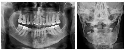
Since glandular odontogenic cysts, as described above, are on the one hand very rare and on the other hand often do not cause any symptoms over a long period of time, they are sometimes only noticed as incidental findings in radiological examinations. If a targeted examination is made for glandular odontogenic cysts, a panoramic tomography (orthopantomogram) is the best method. The cysts show up on the images as sharply delimited lightening that clearly stands out from the bone.
Due to the sometimes aggressive growth of the cysts, dislocations or root resorptions can appear on the adjacent teeth. However, there are no clear pathognomonic radiological signs of glandular odontogenic cysts. In any case, the diagnosis can only be confirmed through a histological investigation. Immunohistochemical markers and the typical glandular tissue of the glandular cysts can be helpful.
Differential diagnoses to be considered include ameloblastoma, odontogenic myxofibroma, central giant cell granuloma, keratocystic odontogenic tumor, follicular cyst, lateral periodontal cyst, and plasmacytoma. Glandular odontogenic cysts can, undetected and therefore untreated, lead to bone damage through osteolysis of the cortex.
Complications
In most cases, the patient does not experience any particular symptoms or complications as a result of the odontogenic cyst. For this reason, this cyst is usually only discovered by chance and treatment is often started late. This can lead to swelling of the jaw.
If the odontogenic cyst continues to grow, asymmetries in the face can occur, which primarily have a negative effect on the patient’s aesthetics and can lead to complications. It is not uncommon for those affected to suffer from feelings of shame or inferiority complexes and the patient’s quality of life is reduced. In the case of a tumor, it can spread to other regions of the body and cause damage and discomfort there.
When treating the odontogenic cyst, in most cases there are no symptoms or complications. It is simple and quickly leads to a positive course of the disease. However, in many cases a new treatment is necessary for the patient and it cannot be ruled out that the odontogenic cyst will reappear at a later point in time. Furthermore, the person concerned is dependent on regular controls. There is generally no reduction in life expectancy.
When should you go to the doctor?
Irregularities in the mouth should be clarified by a doctor. If swellings, ulcers or lumps form in the mouth, a doctor should be consulted. If the person concerned can detect changes in the gums with his tongue, a check-up is recommended.
Since the glandular odontogenic cyst often remains symptom-free and unnoticed for a long time, a doctor should be visited as soon as the first uncertain perceptions are made. If there is a slight sensation of pain in the jaw or a pulling sensation in the mouth when moving the jaw, a doctor should be consulted.
Teeth loosening or shifting are cause for concern. If you feel pressure in your mouth, have trouble cleaning your teeth or have an unusual taste in your mouth, you should see a doctor. If asymmetries in the face or deformities of the face or neck can be noticed, a doctor’s visit is required. If the visual changes result in emotional or psychological complaints, a doctor should be contacted. Persistent feelings of shame or decreased self-esteem should be discussed with a doctor.
Consult a doctor in the event of swallowing difficulties, changes in vocalization or impairments while eating. If you experience discomfort when wearing braces or if you have problems with existing dentures, you should see a dentist as soon as possible.
Treatment & Therapy
Glandular odontogenic cysts can only be treated surgically. Conservative as well as aggressive or resective measures can be found within the surgical options. Conservative suggestions include: cystectomy alone, marsupialization for cysts that are difficult to access, cystectomies or curettages combined with partial peripheral ostectomy.
The combination of cystectomies or curettages with the adjuvant application of Carnoy’s solution, cryotherapy and continuity resections. In the event of an aggressive surgical approach involving resection, reconstruction should be carried out immediately. The method of choice depends on the parameters of the specific case, such as the location, size, and number of the cysts.
Cystectomies can be used for small, individual cysts that only affect one or two adjacent teeth. Multilocular lesions, on the other hand, require more aggressive approaches to prevent recurrences as much as possible. In many cases, the therapy cannot be regarded as complete, as the recurrence rate of up to 35.9 percent often necessitates repeated treatments.
Cases treated by resections show the rarest recurrences. Conservative surgical therapy approaches are made more difficult by the presence of microcysts, and the often extremely thin cyst follicle prevents complete removal.
The risk of recurrence is particularly high in the case of very large and multilocular cysts associated with perforation of the cortex. Regular checks are therefore essential after the surgical treatment. They should be done after three, six and twelve months and continued with annual radiological monitoring.
Outlook & forecast
The prognosis of the glandular odontogenic cyst is to be assessed according to the individual circumstances. In most cases, however, it is documented as favorable.
In some patients, there is no significant impairment or disturbance from the cyst. They are removed conservatively and then the person affected can be discharged from treatment as symptom-free. Although the prognosis is favorable, a new cyst can develop later in life. If it is noticed early and is in a favorable position, the prognosis is good again.
With a difficult localization of the cyst and with an increasing size, the effort of removal increases. In addition, more complications can occur. Shifting of teeth and damage to the bones are possible. Although the removal of the cyst is usually successful, necessary corrections are often advised. In surgical interventions, the teeth are repaired and fastened so that no further sequelae occur. The larger a cyst, the more likely it is to recur.
Although the initial prognosis is favorable, repeated disturbances and new formation of a cyst can occur over the course of the life cycle. For the patient, this means that he should undergo regular check-ups so that a recurrence is noticed as quickly as possible.
Prevention
Since the mechanism of formation of glandular odontogenic cysts is unknown, it is not possible to prevent their occurrence by taking preventive measures. Regular dental checkups, however, increase the likelihood of detecting the cysts early on before they can cause symptoms to the patient. Due to the high risk of recurrence, regular radiological controls are urgently recommended after the diagnosis and treatment of glandular odontogenic cysts has already been carried out.
Aftercare
With this disease there are few or hardly any measures and options for follow-up care available. The patient is primarily dependent on an early diagnosis and detection of this disease so that there is no further compilation or other damage to the bones. Therefore, the early detection and subsequent treatment of this disease is in the foreground in this disease.
Even after a successful treatment, further and regular examinations should be carried out in order to identify and remove other tumors. This cyst may also reduce the life expectancy of the person affected. This disease is treated with the help of various therapies and also by surgical removal of the cysts. The affected person should definitely recover and rest his body after such an operation.
You should refrain from exertion or other physical activities so that the body is not unnecessarily stressed. Regular examinations are necessary in order to detect the recurrence of this complaint at an early stage, even a few years after removal. As a rule, no further follow-up measures are necessary for this disease.
You can do that yourself
Glandular odontogenic cysts often cause no noticeable symptoms for a long time and therefore initially go unnoticed by those affected. However, they run the risk of damaging the bones, so if the patient notices the glandular odontogenic cyst, see a doctor immediately.
In this way, the person affected prevents their quality of life from deteriorating in the long term as a result of the disease. After the doctor removes the glandular odontogenic cyst as part of a surgical procedure, the patient usually suffers from temporary symptoms such as pain and difficulty ingesting food.
First of all, after the operation of the glandular odontogenic cyst, the patient pays particular attention to daily long periods of rest with a lot of sleep or sedentary activities. Because sufficient recovery is important for the regeneration of the body after the stress caused by the operation. The patient spends a lot of time at home and takes care not to expose himself to physical or psychological stress.
If the pain causes problems with eating, the patient adjusts the type of food in consultation with the doctor and prefers soft foods for a certain period of time. Thorough dental and oral hygiene is just as important, as the susceptibility to infections is increased after the operation.